Chi siamo

La struttura di microscopia elettronica del Dipartimento di Scienze della Vita vanta una solida reputazione internazionale per l’utilizzo avanzato di tecniche di preparazione, imaging e post-imaging, applicate a indagini ultrastrutturali su campioni biologici, nanomateriali e studi di morfologia funzionale. Le sue competenze si estendono alla ricerca di base, alla biomedicina, alle biotecnologie e alla scienza dei materiali. La struttura è dotata di un’ampia gamma di strumenti per la preparazione dei campioni, tra cui apparecchiature per il sezionamento ultrasottile, vari sistemi di sputtering e metallizzazione, nonché tecnologie per il congelamento ultrarapido, essenziali per la crio-microscopia elettronica. Per quanto riguarda l’imaging, la dotazione comprende un microscopio elettronico a scansione ambientale (ESEM), ideale per analisi di superficie e determinazione della composizione elementare locale tramite spettrometria EDS. Inoltre, sono disponibili tre microscopi elettronici a trasmissione a media tensione, utilizzabili per indagini a contrasto negativo o per l’analisi di sezioni ultrasottili di campioni inglobati in resina plastica.
Recentemente, la struttura ha arricchito la propria dotazione strumentale con un microscopio elettronico a trasmissione ad alta tensione, dotato di sorgente a emissione di campo (FEG), rivelatore STEM, filtro di energia e spettrometro EDS ad alte prestazioni, consentendo analisi ultrastrutturali ad altissima risoluzione.
Il crioTEM, già operativo da anni, è stato recentemente sostituito con un modello di nuova generazione, anch’esso dotato di sorgente FEG, caricatore automatico per campioni di acqua vetrificata, filtro di energia e rivelatore di elettroni diretti. Lo strumento permetterà l’analisi ultrastrutturale di complessi macromolecolari di rilevanza biologica e biomedica con una risoluzione senza precedenti. Lo staff tecnico della struttura offre servizi di consulenza, imaging e post-imaging su appuntamento, previa valutazione preliminare. Questi servizi sono disponibili per gruppi di ricerca dell’Università di Siena, nonché per ricercatori di altri enti pubblici e privati, compresi progetti commissionati da terzi, secondo il tariffario consultabile su questa pagina web.
Servizi
Il laboratorio di Microscopia Elettronica offre i seguenti servizi:
- Preparazione dei campioni e inclusione in resina
- Ultramicrotomia
- Colorazione negativa
- Immuno-microscopia elettronica
- Sputter coating
- Assistenza nella microscopia elettronica a trasmissione e nell’imaging digitale
- Tomografia elettronica: acquisizione di immagini in serie, allineamento e ricostruzione di modelli derivanti da campioni inclusi in resina
- Consulenza e assistenza nella progettazione di esperimenti di microscopia elettronica
Servizi della struttura di Cryo-Microscopia Elettronica
Questa struttura è una “user facility” e attualmente non fornisce servizi di cryo-microscopia elettronica. Le parti interessate a valutare la compatibilità dei propri campioni per lavori di cryo-microscopia elettronica dovrebbero contattare il direttore della struttura, Prof. Pietro Lupetti
Personale
- P. Lupetti (Direttore del Laboratorio)
- E. Paccagnini (Protocolli di microscopia elettronica, imaging ed elaborazione delle immagini)
- A. Gradi (Manutenzione delle apparecchiature di microscopia elettronica)
- D. Mercati (Protocolli di microscopia elettronica a bassa temperatura)
- L. Masi (Preparazione dei campioni di microscopia elettronica e imaging)
Microscopi Elettronici
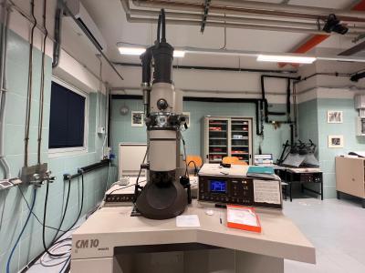
Philips CM10
Il Philips CM-10 TEM è uno strumento stabile adatto sia all’insegnamento universitario che alla ricerca. Questo TEM possiede un porta-campioni goniometrico ed è anche capace di operare in modalità low dose. Il microscopio è semplice da utilizzare e genera immagini di alta qualità e alta risoluzione a voltaggi
convenzionali.
Caratteristiche principali:
- Tensione di accelerazione: 40-100 kV
- Risoluzione (lente obiettivo): 0,5 nm / 5,0 ångström (punto), 0,34 nm / 3,4 ångström (linea)
- Ingrandimento: 20-450.000X
- Telecamera digitale Veleta per l’acquisizione delle immagini
- Modalità operative low dose, campo chiaro e campo scuro

FEI Tecnai G2 Spirit
TEM ad alto contrasto e alta risoluzione, operante a tensioni comprese tra 20 e 120 kV. Adatto per imaging 2D e 3D di sezioni ultrasottili di cellule, organelli cellulari e materiali soffici. Dotato di funzionalità per la tomografia elettronica e la CryoEM di campioni idratati.
Caratteristiche principali:
- Risoluzione: 0,34 nm
- Tensione di accelerazione: 20-120 kV
- Ingrandimento: 22x - 340.000x
- Angolo di inclinazione: +/- 80°
- Telecamera CMOS Tvips bottom-mount 2Kx2K
- Elevato livello di automazione: Auto-Gun e regolazione automatica
- Sistema di posizionamento Smart Tracking per la navigazione del campione
- Osservazione dei campioni alla temperatura dell’azoto liquido
- Tecnologia per un imaging nitido di campioni più spessi
- Software I-TEM

Tecnai F20 FEG
Il Tecnai F20 FEG è un microscopio elettronico a emissione di campo dotato di una lente obiettivo X-TWIN.
Caratteristiche principali:
- Rivelatore EDX Oxford
- Filtro di imaging Gatan Tridiem per spettroscopia EELS
- Risoluzione TEM: 2,4 Å
- Risoluzione STEM: migliore di 2 Å
Fei Quanta 400
Il Fei Quanta 400 ESEM è un microscopio elettronico a scansione ad alta risoluzione.
Caratteristiche principali:
- Risoluzione: 4 nm
- Ingrandimento massimo: 50.000x
- Rivelatore di elettroni secondari
- Rivelatore di elettroni retrodiffusi (Backscattered)
- Rivelatore EDS
- Modalità ESEM: basso vuoto, alto vuoto, modalità umida
- Piattaforma Peltier
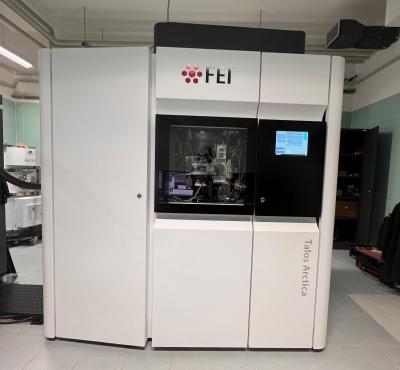
FEI Talos Arctica
Il FEI Talos Arctica è un microscopio elettronico a trasmissione da 200 kV ad alte prestazioni, completamente automatizzato, progettato specificamente per la crio-microscopia elettronica.
Caratteristiche principali
- X-FEG da 200 kV
- Digital FluCam – consente esecuzione manuale e automatica degli allineamenti durante la ricerca e la visualizzazione
- Telecamera Ceta 16M
- Autoloader – capacità di caricare fino a 12 griglie simultaneamente in condizioni criogeniche
- Raccolta dati automatizzata
- Telecamera digitale Gatan K3
- Filtro Bio Quantum GIF
STRUMENTAZIONE PER LA PREPARAZIONE DEI CAMPIONI
Sistemi di frattura e incisione a freddo:
- Balzer’s BAF 400
- Balzer’s BAF 301
Dispositivi per il congelamento ultrarapido:
- FEI Vitrobot MK4
- Leica EM GP2 Automatic Plunge Freezer
- MedVac Cryopress
- Reichert Plunge Freezer
- Balzer’s FSU Freeze Substitution Unit
Ultramicrotomi:
- Reichert Ultracut
- Reichert Ultracut II E
- LKB Nova
Preparazione per SEM:
- Balzer’s Med 010 Mini Deposition System
- Balzer’s CpD 010 Critical Point Dryer
- Edwards Gold Sputter Device