
The technological platform for electron microscopy at the Department of Life Sciences has a well-established and internationally recognized reputation for the use of various preparative, imaging, and post-imaging techniques for ultrastructural investigations on biological samples, nanomaterials, and functional morphology studies in basic research, biomedicine, biotechnology, and materials science.
The platform is equipped with all the necessary instrumentation for sample preparation, ultrathin sectioning, various sputtering and metallization methods, and ultrarapid freezing for cryo-electron microscopy investigations.
Regarding imaging, the platform has an ESEM for surface investigations and local elemental composition determination of samples using EDS spectrometry.
Additionally, three electron microscopes with medium acceleration voltage are available for negative contrast investigations or imaging of ultrathin sections from plastic embedded samples.
A high-voltage transmission electron microscope for high-resolution ultrastructural analyses has also been recently installed, equipped with a field emission gun (FEG), STEM detector, energy filter, and high performance EDS spectrometer.
The cryoTEM, operational at the platform for many years is being replaced with a next-generation cryoTEM featuring a FEG source, autoloader for vitrified water samples, energy filter, and direct electron detector. The instrument will enable ultra-high-resolution ultrastructural investigations of macromolecular complexes of biological and biomedical interest.
The platform's technical staff provides consulting, imaging, and post-imaging services by appointment and after an exploratory meeting, for research groups within the University of Siena as well as for researchers from other public and private research institutions, including third-party projects, according to the pricing published on this web page
Electron Microscopy facility provides the following services:
- Sample processing, embedding in plastic resin
- Ultramicrotomy of plastic sections
- Negative staining
- Immuno-electron microscopy
- Sputter coating
- Assisting in transmission electron microscopy and digital imaging
- Electron tomography: tilt series acquisition, image alignment and reconstruction for plastic embedded samples
- Consultation and assistance in design of EM experiments
Cryo EM facility service
This facility is a "user facility" and currently does not provide cryo EM service. Any parties interested in having samples evaluated for suitability for cyro EM work should contact EM facility director, Prof. Pietro Lupetti
P. Lupetti (Lab. Director)
E. Paccagnini (EM protocols, imaging and image processing)
A. Gradi (EM equipments maintenance)
D. Mercati (Low temperature EM protocols)
L. Masi (EM sample processing and imaging)
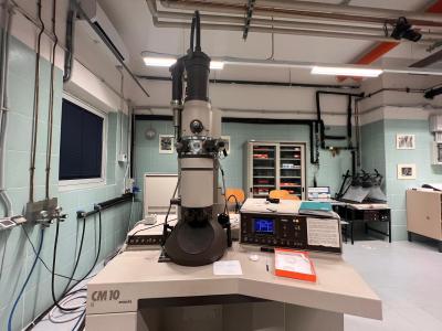
Philips CM10
The Philips CM-10 TEM is a stable instrument suited to both undergraduate teaching and research. This TEM possesses a goniometer specimen stage, and is also capable of low dose operation. The microscope is simple to operate and generates high quality, high resolution images at conventional voltages.
Main features
Accelerating voltage: 40-100kV
Resolution (objective lens): 0.5 nm/5.0 ångströms (point), 0.34 nm/3.4 ångströms (line)
Magnification: 20- 450,000X
Veleta digital camera for image acquisition
Low dose, bright & dark field operation modes

FEI Tecnai G2 Spirit
High contrast, high resolution TEM for 20 –120 kV operation. Suitable for 2D & 3D imaging of thin sectioned cells, cell organelles and soft matter. Also fitted for Electron tomography and CryoEM of hydrated samples
Main features

Tecnai F20 FEG
The Tecnai F20 FEG is a field emission gun electron microscope equipped with an X-TWIN objective lens.
Main features
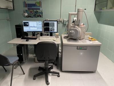
Fei Quanta 400
The Fei Quanta 400 ESEM is a high resolution scanning electron microscope.
Main features
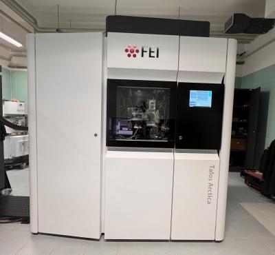
- 200kV X-FEG
- Digital FluCam – All manual and automatic alignments can be executed with the search and view
- Ceta 16M Camera
- Autoloader - up to 12 grids can be loaded simultaneously under cryogenic conditions
- Automated data collection
- Gatan K3 digital camera
- Bio Quantum GIF Filter
Freeze Fracture & Freeze Etch Systems:
Ultrarapid Freezing Devices:
Ultramicrotomes:
SEM Processing: